Collagen is the second most abundant substance in the human body (immediately after the water), and represents about 30% of the total protein. Provides structural support for virtually all organs and soft tissues (including joints). Unfortunately, as we age, our body tends to produce less and less collagen. In fact, upon reaching 60 years of age, the ability to produce collagen is reduced by 50%, thus causing aging of the joints, loss of skin elasticity and loss of lean mass. From here it is clear that the preservation of a good mass of collagen in the joints and skin is an integral part of healthy aging.

Joints and muscles are tied together by connective tissue. In addition to providing structural integrity, connective tissue helps transmit strength, protect muscles and bones from injury, transport nutrients, and repair damaged cells. Each structure plays a different role in the muscle strength production process, which depends not only on the anatomical structure but also on the content and arrangement of the collagen Fibre.
In fact, there are different types of joint connective tissue:
- Tendons: Tendons connect muscles to bones. They are more elastic than ligaments but stiffer than muscles. They are responsible for much of the transfer of force through the body during movement.
- Ligaments: Ligaments connect bones to other bones. Their main purpose is to stabilize.
- Cartilage: Cartilage acts as a cushioning structure within the joints and between the bones. Unlike tendons and ligaments, cartilage lacks blood vessels and nerves, making it a problematic type of connective tissue for the body to repair.
- Fascia: fascia is the thin sheath of connective tissue that surrounds muscles. It plays a vital role in the transfer of force between the parts of the kinetic chain. The fascia is dense with nerve endings, making it almost as sensitive as the skin. This is part of the reason why manual therapy methods (such as foam rolling and massage) are effective for pain and tension relief. Over 80% of the muscle force produced is transferred to the surrounding connective tissue.
Intramuscular connective tissue, tendons, ligaments, and the fascia sheath that surrounds muscles are all composed primarily of collagen. In particular, from type I collagen.
In fact, there are different types of collagen (28 have been described in the literature), with specific functions and characteristics. Among the best known is type I collagen (which represents 90% of total collagen and constitutes, precisely, the main connective tissues), type II collagen (fundamental to cartilage tissue), type III (present in the dermis and blood vessels) and type IV (with support functions and component of the basement membrane).

During the recovery process from training, environmental variables such as movement habits, nutrition quality and inflammation levels can help or hinder this repair process. The two main influencing factors are the total collagen available for cross-linking and the quality of collagen fibril formation. In terms of controllable factors, exercise has the most significant positive impact on collagen synthesis within the joints. Despite this, several lifestyle variables matter.
What to avoid (or better still use with caution) and what to pay attention to:
1) NSAIDs (Non-Steroidal Anti-Inflammatory Drugs), such as ibuprofen and naproxen, reduce the mass of collagen at the injury sites. A metastude published in the Annals of Physical and Rehabilitation Medicine showed that although NSAIDs are able to effectively relieve joint pain and reduce inflammation in the short term (7 to 14 days), they also delay healing times, they also increase re-injury rates of up to 25% and also reduce the mass of collagen in the injured sites. Corticosteroids also help relieve pain and inflammation in the short term, but have inhibitory effects on collagen synthesis within connective tissue.
2) A sedentary lifestyle leads to a decrease in total collagen in the body, while exercise focused on using overloads increases the rate of collagen formation. During periods of injury recovery, a reduction in activity leads to a reduction in collagen, further increasing the risk of injury.
3) Age. Collagen production decreases with age. By the age of 60, the ability to produce collagen decreased by approximately 50%.
4) An imbalance of testosterone and estrogen can inhibit collagen synthesis. Too much estrogen also has a detrimental effect on collagen health by decreasing its stiffness, making it easier for connective tissue to break.
Supplements to improve collagen
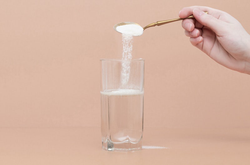
Before moving on to a list of supplements that have been scientifically proven to support the production of endogenous collagen, we remind you that it is possible to directly take supplements based on hydrolyzed collagen. Collagen is one of the most important structural Protein present in our organism, where it represents about 1/3 of the total Protein; as explained above, it is also the main constituent of connective tissue (80%), the tissue that makes up skin, muscles, hair, nails and cartilage. Of the total, 40% of collagen is contained in the skin, where it guarantees elasticity and firmness. By taking hydrolyzed collagen, a highly bioavailable form (that is, easily usable by our body) we will effectively supply precious bricks to restructure and recompact our skin, our joints and our tissues in general, giving greater vigor, resistance, elasticity and turgor.

Below is a list of supplements that have been shown to improve and support collagen production, quantity and quality.
Whey Protein: A study published in the Scandinavian Journal of Medicine and Science in Sports showed that subjects who used whey protein isolate in combination with resistance training saw a greater increase in tendon mass. A consumption of at least 20-40 grams per day is recommended, ideally within an hour of exercising.
Essential Amino Acids (EAAs): While protein supplementation has more research support than using amino acid supplements, EAAs can still provide the same or better results than whey protein. Studies have shown that EAA supplementation stimulates muscle protein synthesis (MPS) more than the ever popular branched chain amino acid (BCAA) supplement, and both MPS and whey protein do. Unlike the latter, EAAs have the added benefit of creating lower insulin spikes and easier digestion.
Vitamin C + Collagen Protein: An eight-person placebo-controlled study published in the American Journal of Clinical Nutrition showed that taking vitamin C along with collagen protein can double the markers of collagen synthesis in the joints of the ankle. Collagen protein and vitamin C supplements taken individually also have regenerative benefits. A study published in the Journal of Sports Science and Medicineshowed that 5 grams of collagen peptides significantly improved the perceived function of injured ankle joints and decreased the risk of new ankle injury after a three-month follow-up. A metastudio that has collected over 60 scientific studies also concluded that supplementation with collagen is effective, and stimulates the regeneration of connective tissue by increasing not only the synthesis of collagen but also that of minor components (glycosaminoglycans and hyaluronic acid).
Type II collagen: A study published in HealthMED showed that supplementing with 750mg of a natural collagen matrix composed of 93% type II collagen stimulates collagen synthesis within cartilage tissues. Isolated type II collagen supplements with dosages starting at 10-40 mg per day have also shown benefits for joint pain and inflammation.
Bibliography
Lugo, J. P., Saiyed, Z. M., Lau, F. C., Molina, J. P., Pakdaman, M. N., Shamie, A. N., & Udani, J. K. (2013). Undenatured type II collagen (UC-II®) for joint support: A randomized, double-blind, placebo-controlled study in healthy volunteers. Journal of the International Society of Sports Nutrition, 10(1), 48. https://doi.org/10.1186/1550-2783-10-48
Bakilan,F.,Armagan,O.,Ozgen,M.,Tascioglu,F.,Bolluk,O.,&Alatas,O.(2016).Effects of native type II collagen treatment on knee osteoarthritis: A randomized controlled trial. The Eurasian Journal of Medicine, 48(2), 95–101. https://doi.org/10.5152/eurasianjmed.2015.15030
Figueres Juher, T., & Basés Pérez, E. (2015). Review of the beneficial effects of hydrolyzed collagen intake on osteoarticular health and skin aging [An overview of the beneficial effects of hydrolyzed collagen intake on joint and bone health and on skin ageing]. Hospital Nutrition, 32 Suppl 1, 62–66. https://doi.org/10.3305/nh.2015.32.sup1.9482
Dressler,P.,Gehring,D.,Zdzieblik,D.,Oesser,S.,Gollhofer,A.,&König,D.(2018). Improvement of functional ankle properties following supplementation with specific collagen peptides in athletes with chronic ankle instability. Journal of Sports Science & Medicine,
Shaw,G.,Lee-Barthel,A.,Ross,M.L.,Wang,B.,&Baar,K.(2017).VitaminC-enriched gelatin supplementation before intermittent activity augments collagen synthesis. The American Journal of Clinical Nutrition, 105(1), 136–143. https://doi.org/ 10.3945/ajcn.116.138594
Park,S.,Church,D.D.,Azhar,G.etal.(2020).Anabolicresponsetoessentialaminoacid plus whey protein composition is greater than whey protein alone in young healthy adults. J Int Soc Sports Nutr, 17, 9 (2020). https://doi.org/10.1186/s12970-020-0340-5
Varani,J.,Dame,M.K.,Rittie,L.,Fligiel,S.E.,Kang,S.,Fisher,G.J.,&Voorhees,J.J. (2006). Decreased collagen production in chronologically aged skin: Roles of age-dependent alteration in fibroblast function and defective mechanical stimulation. The American Journal of Pathology, 168(6), 1861–1868. https://doi.org/10.2353/ajpath.2006.051302
Ramaswamy,K.S.,Palmer,M.L.,vanderMeulen,J.H.,Renoux,A.,Kostrominova,T.Y., Michele, D. E., & Faulkner, J. A. (2011). Lateral transmission of force is impaired in skeletal muscles of dystrophic mice and very old rats. The Journal of Physiology, 589(Pt 5), 1195– 1208. https://doi.org/10.1113/jphysiol.2010.201921
Leitman,D.C.,Benson,S.C.,&Johnson,L.K.(1984).Glucocorticoidsstimulatecollagen and noncollagen protein synthesis in cultured vascular smooth muscle cells. Journal of Cell Biology,
98, 541–549. https://rupress.org/jcb/article pdf/98/2/541/1390360/541.pdf


Comments
Write a comment about the article